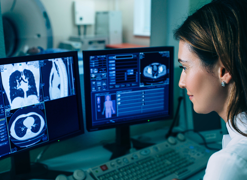
 The curriculum of the Cardiothoracic Imaging fellowship includes thoracic, cardiac, interventional and elective rotations as well as time for research and participation in interdisciplinary pulmonary and thoracic oncology conferences.
Excellent working relations are maintained with:
- Pulmonary medicine
- Cardiothoracic surgery
- Hematology-oncology
- Cardiology
- Pathology
Technology and Equipment
The Cardiothoracic Imaging fellowship program has access to:
- Nine fast reconstructing multi-detector computed tomography scanners
- Seven state-of-the-art ultrasound units
- Five MR imaging units
- 1.5T MR unit dedicated to cardiovascular imaging
- Six remote control radiographic/fluoroscopic rooms
-
Special procedure rooms for:
- visceral arteriography
- digital subtraction angiography
- ultrasound
- interventional procedures
Overall Program Objectives
- Train fellows to become experts in cardiothoracic imaging across multiple modalities (CT, MRI, X-ray, nuclear medicine, and ultrasound).
- Foster skills in diagnosis, reporting, and clinical consultation with a focus on thoracic and cardiovascular diseases.
- Encourage academic development through research, presentations, and teaching opportunities.
- Promote multidisciplinary collaboration with pulmonology, cardiology, and oncology departments.
