Brachytherapy is a type of internal radiation therapy that delivers a highly focused radiation dose directly to a tumor while minimizing radiation exposure to surrounding healthy tissue. During treatment, tiny radioactive seeds are implanted through the channels of applicators at designated locations to destroy cancer cells. Because of its precise targeting, reduced side effects, and short treatment duration, brachytherapy is widely used for treating localized tumors, including those in the prostate, breast, cervix, and head and neck regions.
For brachytherapy to be successful, precise planning of the locations for applicators and radioactive seeds as well as the radiation dose is critical. This technology is a novel 3D-printed brachytherapy contrast marker that improves visualization of applicators during both CT and MRI-guided treatment planning while minimizing the imaging artifacts commonly caused by conventional metallic markers. By utilizing metal-infused PLA and an optional contrast-filled hollow design, it provides enhanced image contrast and more accurate anatomical mapping for radiation dose planning. This technology offers a low-cost, dual-modality imaging solution that can improve treatment precision, optimize dose delivery, and ultimately enhance patient outcomes in brachytherapy.
Researchers and commercial partners interested in advancing this technology should contact the university's technology transfer office to discuss licensing opportunities, as the university is seeking to partner with qualified entities to scale the manufacturing process and distribute a new global product for brachytherapy treatments.
Technology Overview
Current Challenges
Accurate treatment planning of locations for applicators and radioactive seeds, as well as radiation doses are essential for brachytherapy success. It is the gold standard to integrate CT and MRI imaging during planning; however, conventional metallic wires used for aiding applicator visualization during CT and or MRI, while at low cost to medical centers, introduce substantial artifacts that interfere with anatomical mapping and dose planning. Accordingly, there is a need for improved low-cost contrast markers that are compatible with both CT and MRI, which minimize artifacts to ensure precise treatment delivery and optimized patient outcomes.
Our Innovation
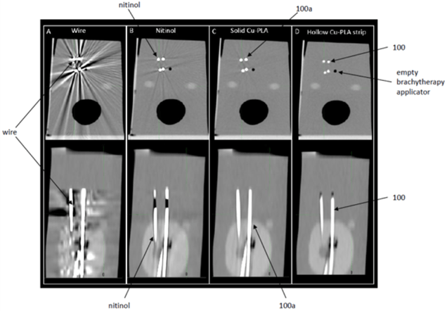
Our novel 3D-printed brachytherapy contrast marker provides an effective solution for brachytherapy planning that is fully compatible with both CT and MRI imaging. By adopting metal-infused polylactic-acid (PLA) filament, the novel 3-D-prined contrast marker has the benefit of enhanced visibility, reduced imaging artifact, and superior CT and MRI contrast, when compared to the commercially available steel or nitinol wires. The physical parameters of the contrast marker are designed to match brachytherapy needles. The contrast marker can be solid or partially hollow, with the hollow part capable of containing viscous contrast media to further enhance imaging contrasts and minimize artifacts.
Benefits of this Technology
The 3D-printed contrast markers improve the precision, efficiency, and cost-effectiveness of brachytherapy treatment planning by providing:
- Enhanced imaging performance: Clear visualization under both CT and MRI with minimal artifact.
- Improved treatment accuracy: Enables precise mapping of applicator channels for optimized dose delivery.
- Customization flexibility: Easily tailored in shape, composition, and length using accessible 3D-printing tools.
- Streamlined workflow: Rapid, in-house customization reduces lead time.
- Cost efficiency: Primary cost limit to 3D-printing filament, which continues to decrease over time.
Stage of Development
The 3D-printed brachytherapy marker has been prototyped and bench tested successfully.
Figures
Figure 1: shows cross-sectional and side views of CT images comparing CT imaging artifacts generated by commercially available contrast markers and the 3D-printed contrast markers (solid & hollow).